Capturing the Unseen World with Microscopic Photography
Microscopic photography falls under many names, including “microscope photography”, “micrography”, and “microscopy”. The microscope is primarily a scientific apparatus designed to magnify tiny objects that are usually invisible to the naked eye. Imagine the power of a microscope used to capture images that may never before have been seen by the average person.
Microscopes Used in Microscopic Photography
Optical and electron microscopes are often used in microscopic photography. Scanning electron microscopes (SEM) are huge contraptions that work by scanning the image of the subject using high-energy electron beams. They can capture fine detailed images using high resolution and a very large depth of field which makes the samples look three-dimensional. Their magnification capacity is also immense.
The optical microscope is the more familiar type that we see in science class. It is also called “light microscope” and makes use of visible light and lenses to magnify small samples. These magnified images, or “micrographs”, can be captured by certain regular cameras. Previously, Single-Lens Reflex (SLR) cameras and photographic film were used to record images. Digital technology has made microscopic photography more accessible and convenient. Digital cameras can be used with microscopes and digital microscopes provide instant gratification by allowing us to view the magnified sample details directly on a computer screen.
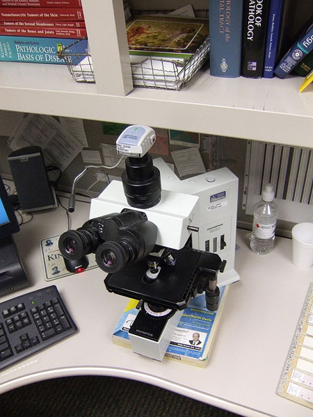
Photo by joebeone
Revealing Details with Microscopic Photography
Through micrography, we can record images of the microscopic world around us which we do not notice. Whenever we feel all alone in the world, just remember all the hundreds of thousands of bacteria and microorganisms all around us. Images of these can be taken in extraordinary detail through microscopic photography. For example, take a look at the micrograph of this flour beetle (approx ¼-1/8 in size). Its fine hairs and segmented body can clearly be seen.
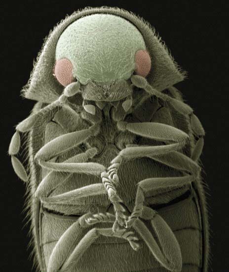
Photo by dullhunk
For those who want an even more magnified sample, how about a highly detailed view of the virus, Salmonella typhimurium (in red), as it invades some cultured human cells. Bear in mind that micrographs are often color-enhanced to make the details even more defined.
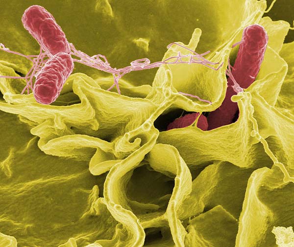
Photo by Nutloaf
Microscopic Photography Can Produce Art Images
Although microscopic photography is greatly beneficial to the field of science, it has also become quite popular in photographic art. After all, seldom can we see such fascinating textures and details of common objects that look so different when viewed up close, really close. Common household objects can be seen in a vastly different light when placed under the focus of a microscope. In this image that shows such stunning texture, who would guess that it is a sample of ordinary torn white bread?
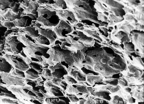
Photo by CORE-Materials
This next photo is gorgeous with its vibrant display of color and patterns. We could call it an abstract photo which is certainly eye-catching. If we found out that the subject used in this photo is of bladder sand (the particles that can accumulate and form into bladder stones), it could give us a different perspective and a deeper appreciation of the world, not just around us, but also within.
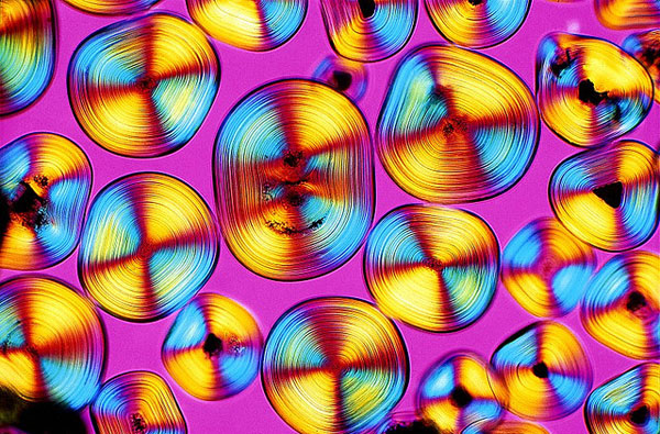
Photo by dullhunk


wonderful Images…
Salmonella typhimurium is a bacteria, not a virus. Two completely different organisms.